
A history of recurrent heartburn along with a positive response to antacids or acid-suppressing medication is adequate to diagnose Acid Reflux Disease. Specific testing is reserved for patients who have (1) Acid Reflux Disease plus alarm symptoms of dysphagia, weight loss, or gastrointestinal bleeding; (2) Acid Reflux Disease of sufficient chronicity (e.g., 5 years) to raise concern for Barrett's esophagus; and (3) suspected Acid Reflux Disease with atypical symptoms, such as chest pain or oropharyngeal, laryngeal, or airway symptoms.
Establishing Acid Reflux Disease as a Cause of Nonheartburn Symptoms
Currently, the preferred method for establishing Acid Reflux Disease as the cause of symptoms (e.g., chest pain, wheezing) is an empirical trial of acid suppression with a PPI (e.g., omeprazole, 20 mg twice daily), which normalizes esophageal acidity in approximately 95% of subjects. In some instances, a bedtime dose of a histamine H2-receptor antagonist (e.g., ranitidine, 300 mg) is added to reduce the possibility of nocturnal acid breakthrough. The treatment period in which to expect a satisfactory response is 2 to 4 weeks for chest pain and 2 to 3 months for inflammatory disease of the airway. Resolution of the symptoms supports Acid Reflux Disease as possibly causal. Confirmation may be obtained by relapse when medication is withdrawn and by a subsequent positive response to re-treatment, as confirmed by documenting control of esophageal acidity on pH monitoring while undergoing PPI therapy. Failure of symptoms to improve with PPI therapy is not generally an indication for antireflux surgery (see later) but rather an indication to search for another disease. A rarely used alternative is the Bernstein test, in which acid (0.1 N HCl, pH 1.1) or saline (control) is perfused through a catheter positioned in the midesophagus. If symptoms typical of those that occur spontaneously develop with acid but not saline, the test is considered positive for Acid Reflux Disease.
Tests for Reflux
Documenting acid reflux is not necessary except when symptoms fail to respond to PPI therapy or when surgery is being considered. Esophageal pH monitoring, the “gold standard” for identifying acid reflux, is performed by fixing a small pH probe in the esophagus, 5 cm above the LES, and recording all episodes in which esophageal pH drops to less than 4 over a 24- to 48-hour period. The number and duration of each acidic event, when combined, yield a value for total esophageal acid contact time. Total acid contact times of greater than 5% are abnormal and consistent with a diagnosis of Acid Reflux Disease. An event marker activated by the patient also allows symptoms to be related to episodes of esophageal acidity. An upper gastrointestinal series can detect grossly abnormal reflux by observing movement of barium from the stomach to the esophagus with the patient in the head-down position. It has low sensitivity but, when positive, has high predictive value. The positive predictive value is much lower, however, if reflux is induced by having the subject sip water through a straw in the head-down position. This test is rarely useful for therapeutic decisions.
Tests for Esophageal Injury
Endoscopic signs.
Tests of Esophageal Motor Function
An upper gastrointestinal series or barium swallow is valuable for identifying gross reflux and marked abnormalities in esophageal anatomy (e.g., hiatal hernia, diverticulum) and peristaltic and sphincter function. More subtle abnormalities, however, require esophageal manometry. Low mean LES pressure (<10 mm Hg) is a specific but insensitive marker of Acid Reflux Disease, with 60% of patients having normal values. Currently, the major uses of esophageal manometry in Acid Reflux Disease are to (1) position the pH probe for reflux testing, (2) exclude motor disease (achalasia, scleroderma), and (3) quantify peristaltic amplitudes before surgical fundoplication. If contraction amplitudes average less than 30 mm Hg, a partial (Toupet) rather than complete (Nissen) wrap may be preferable to avoid postoperative dysphagia.
The history and clinical manifestations of Acid Reflux Disease are the most important diagnostic aids; objective testing can quantify the extent and severity of the process. In the vast majority of sufferers, typical symptoms of Acid Reflux Disease and the response to initial gastric acid suppressive therapy make the diagnosis relatively easy. Diagnostic evaluation becomes important when symptoms are atypical and/or do not respond to therapy.
DOCUMENTING REFLUX.
Reflux during a barium swallow in adults is uncommon unless vigorous provocative maneuvers are employed. When spontaneous reflux of barium is seen, it usually denotes free reflux. The absence of reflux seen radiographically does not, however, imply that the sufferer does not have Acid Reflux Disease.
The 24-hour monitoring of esophageal pH can be performed with a portable unit, which allows the sufferer to follow an almost normal lifestyle. During the prolonged monitoring period, the relationship between symptoms (heartburn, chest pain, wheezing) and episodes of acid reflux can be ascertained, and calculations can be made of the number of episodes of reflux and the amount of time the esophagus is acidified (pH - 4). A small amount of reflux, especially in the postprandial period, can be seen normally. Repeated and prolonged bursts of acid exposure suggest that abnormal gastroesophageal reflux is present.
In children and infants, reflux can be measured noninvasively by scanning the esophageal area with a gamma-camera after placing a solution of 99m Tc sulfur colloid in the stomach. An abdominal binder is used to increase intra-abdominal pressure and to stress the gastroesophageal junction if free reflux is not seen.
LINKING REFLUX TO SYMPTOMS.
If pain is the predominant symptom, rather than heartburn, a Bernstein test may be performed using the same catheter as is used for esophageal manometry. After a 5-minute period of dripping normal saline in the mid-esophagus, the infusion is changed to 0.1 N hydrochloric acid. Reproduction of the symptoms during acid infusion (usually 4 to 5 minutes into the infusion), followed by rapid symptom disappearance after returning to a saline infusion, suggests an esophageal cause of the discomfort.
As another approach, the sufferer is asked to signal the time of discomfort during prolonged pH monitoring of the esophagus. If the sufferer signals discomfort at the same time that acid reflux is demonstrated by the pH probe, then a causal relationship is more likely.
ASSESSING THE EFFECT OF REFLUX ON THE ESOPHAGEAL MUCOSA.
A barium swallow detects gross changes, such as stricture formation or a deep esophageal ulcer, but misses the much more common shallow ulcerations and erosions, which are detected by endoscopy. Only discrete lesions such as erosions and ulcerations should be taken as proof of esophageal damage, because endoscopic findings, such as erythema, edema, or friability, are subject to wide interobserver variation. In approximately one-half of sufferers with moderate to severe symptoms of Acid Reflux Disease, the mucosa appears absolutely normal, but a biopsy may demonstrate the histologic changes of reflux.
APPROACH
Endoscopy is generally indicated if symptoms are prolonged and do not respond to empiric treatment, or if systemic manifestations, such as weight loss, anemia, and occult blood-positive stool are present. If the appearance of the esophageal mucosa is normal during endoscopy, biopsies can also be obtained to search for objective evidence of microscopic esophagitis. If dysphagia is present, a barium swallow is appropriate. Uncommonly, reflux is demonstrated, a stricture found, or a deep ulcer seen, which leads to immediate endoscopy for more complete evaluation. After first evaluation, it may be appropriate to begin empiric therapy (see Treatment, below). If the response to therapy is poor, esophageal pH monitoring can confirm the diagnosis. At the same time, esophageal manometry may be performed to estimate LES pressure and to determine the presence or absence of peristaltic waves.
The most thoroughly evaluated classification scheme for esophagitis is the Los Angeles system, which categorizes mucosal injury as grade A, B, C, or D. However, it is important to note that the Los Angeles system does not consider strictures, hiatal hernia, or Barretts metaplasia; the endoscopist is required to describe these separately.
Los Angeles Endoscopic Grading Scheme for Esophagitis Severity
Grade A
One (or more) mucosal breaks no longer than 5 mm that do not extend between the tops of two mucosal folds.
Grade B
One (or more) mucosal breaks more than 5 mm long that do not extend between the tops of two mucosal folds.
Grade C
One (or more) mucosal breaks that are continuous between the tops of two or more mucosal folds but involve lesser than 75% of the circumference.
Grade D
One (or more) mucosal breaks that involve at least 75% of the esophageal circumference.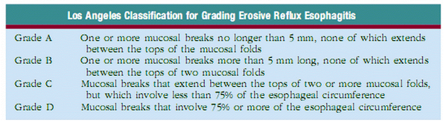


DIAGNOSTIC EVALUATION
Many tests are available for evaluating patients with suspected GERD. These tests are often unnecessary because the classical symptoms of heartburn and acid regurgitation are sufficiently specific to identify reflux disease and to begin medical treatment. However, this may not always be the case, and the clinician must decide which test to choose to arrive at a diagnosis in a reliable, timely, and cost-effective manner, depending on the information desired.
The simplest and most definitive method for diagnosing GERD and assessing its relation to symptoms (either classical or atypical) is the empiric trial of acid suppression. Unlike other tests that only suggest an association (e.g., esophagitis at endoscopy or positive symptom index on pH testing), the response to antireflux therapy ensures a cause-and-effect relationship between GERD and symptoms. Therefore, it has become the “first” test used in patients with classical or atypical reflux symptoms without “alarm” complaints. The popularity of this approach was aided by the introduction of the PPIs, which, unlike the histamine 2 receptor antagonists (H 2RAs), could drastically reduce the amount of acid reflux into the esophagus. Symptoms usually respond to a PPI trial in 7 to 14 days. If symptoms disappear with therapy and then return when the medication is stopped, GERD may be assumed.
In the reported empiric trials with heartburn, the initial dose of PPI was high (e.g., omeprazole 40 to 80 mg/d) and was given for not less than 14 days. A positive response is defined as at least 50% improvement in heartburn. Using this approach, the PPI empiric trial had a sensitivity of 68% to 83% for determining the presence of GERD. Empiric trials using a 2- to 4-month regimen of PPIs taken twice a day also are commonly used in patients with suspected GERD-associated asthma and GERD complaints related to the ear, nose and throat.
An empiric trial of PPIs for diagnosing GERD has many advantages. The test is office based, is easily performed, is relatively inexpensive, is available to all physicians, and avoids many needless procedures. Disadvantages are few, including a placebo response and uncertain symptomatic end point if symptoms do not resolve totally with extended treatment.
Esophageal Manometry
Esophageal manometry allows accurate assessment of LES pressure and relaxation, as well as peristaltic activity including contraction amplitude, duration, and velocity. However, esophageal manometry is generally not indicated in the evaluation of the patient with uncomplicated GERD because most of these patients have a normal resting LES pressure. It is an integral component of pH testing to define the LES location accurately, a task poorly performed by endoscopy, fluoroscopy, or the pH pull-through technique. Esophageal manometry is an essential test in the preoperative evaluation of patients for antireflux surgery. A normal LES pressure does not preclude surgery for the reasons discussed, yet occasionally an alternative diagnosis such as achalasia or scleroderma is made, which may change the clinical approach. Most importantly, the presence of ineffective peristalsis characterized by either low-amplitude (<30>
Bile reflux can be measured using ambulatory esophageal bilirubin monitoring (Bilitec: Medtronics,
Symptoms associated with GERD may be mimicked by other esophageal and extraesophageal diseases including achalasia, Zenker diverticulum, gastroparesis, gallstones, peptic ulcer disease, functional dyspepsia, and angina pectoris. These disorders usually can be identified by failure to respond to aggressive antisecretory therapy and by diagnostic tests such as endoscopy, barium esophagram, esophageal manometry, ultrasound, nuclear emptying studies, and various cardiac tests. Although GERD is the most common cause of esophagitis, other causes (esophagitis, infections, or radiation esophagitis) need to be considered in cases that are difficult to manage cases and in older or immunocompromised patients.
Barium Esophagram
The barium esophagram is an inexpensive, readily available, and noninvasive esophageal test. It is most useful in demonstrating structural narrowing of the esophagus and in assessing the presence and reducibility of a hiatal hernia. Subtle findings such as Schatzki rings, webs, or minimally narrowed peptic strictures are often seen only with an esophagram; they are missed by endoscopy, which may not adequately distend the esophagus. This test, which involves consuming a 13-mm radiopaque pill or marshmallow along with the barium liquid, is the most sensitive for detecting esophageal narrowing, with values reported between 95% and 100%. By giving the patient in the prone oblique position swallows of barium, the barium esophagram also allows good assessment of peristalsis and is helpful preoperatively in identifying a weak esophageal pump.
The ability of the barium esophagram to detect esophagitis varies considerably. Although sensitivities of 79% to 100% have been reported with moderate to severe esophagitis, mild esophagitis is usually missed. Barium testing also falls short when addressing the presence of Barrett esophagus. Barium studies can identify GER when contrast moves in a retrograde fashion from the stomach into the esophagus. If this occurs spontaneously, repeatedly, or to a significant degree into the middle or proximal esophagus, the test is positive, but it has a sensitivity of only about 40% for defining GERD. Provocative maneuvers such as leg lifting, coughing, the Valsalva maneuver, or the water-siphon test can be used to elicit stress reflux. Although these tests can improve the sensitivity of the barium esophagram, some argue that they also decrease its specificity.
The barium esophagram is primarily used in evaluating the patient with GERD with new-onset dysphagia because it can define subtle strictures and rings as well as assess motility. Conversely, endoscopy is preferred in the patient with recurrent dysphagia known to have a stricture or for the assessment of esophagitis or Barrett esophagus.
Esophageal pH Monitoring
Ambulatory intraesophageal pH monitoring is now the standard for establishing pathological reflux. The test is performed with a pH probe passed nasally and positioned 5 cm above the manometrically determined LES. The probe is connected to a battery-powered data logger capable of collecting pH values every 4 to 6 seconds. An event marker is activated by the subject in response to symptoms, meals, and body position changes. Patients are encouraged to eat normally and to pursue regular daily activities. Monitoring is carried out usually for 18 to 24 hours. Reflux episodes are detected by a drop in pH to less than 4. Commonly measured parameters include the percentage of total time that the pH is less than 4, the percentage of time upright and supine that the pH is less than 4, the total number of reflux episodes, the duration of longest reflux episode, and the number of episodes longer than 5 minutes. The total percentage of time that the pH is less than 4 is the most reproducible measurement for GERD, with reported upper limits of normal values ranging from 4% to 5.5%. Ambulatory pH testing can discern positional variations in GER, meals, and sleep-related episodes and helps to relate symptoms to reflux events. As the result of its reliability for measuring GER across normal activities, ambulatory pH testing has replaced other older studies, such as the standard acid reflux (Tuttle) test and radionuclide scintigraphy.
One important problem with esophageal pH monitoring is that there exists no absolute threshold value that reliably identifies pathological GER. Validation studies comparing the presence of esophagitis with abnormal pH test report sensitivities ranging from 77% to 100% with specificities from 85% to 100%. However, these patients rarely need pH testing; rather, the patients with normal endoscopic findings and suspected reflux symptoms should benefit most from ambulatory pH monitoring. Unfortunately, the data are much less conclusive in this group, with considerable overlap between controls and patients with nonerosive reflux. Other drawbacks of pH testing include possible equipment failure, the pH probe’s missing a reflux event because it is buried in a mucosal fold, and false-negative studies resulting from dietary or activity limitations from poor tolerability of the nasal probe.
An important advantage of ambulatory esophageal pH monitoring is its ability to record and correlate symptoms with reflux episodes over extended periods. For this indication, it has essentially replaced the shorter acid perfusion (Bernstein) test. Because only about 10% to 20% of reflux episodes are associated with reported symptoms, different statistical analyses have evolved attempting to define a significant association between these two variables including the symptom index, symptom sensitivity index, and symptom association probability. Unfortunately, no studies to date have defined the accuracy of any of these symptom scores in predicting response to therapy. Therefore, pH testing and symptom correlation can define an association between complaints and GER, but only treatment trials address the true definition of a causal relationship.
Definite clinical indications for ambulatory pH monitoring have been established. Before fundoplication, pH testing should be performed in patients with normal endoscopic findings to identify the presence of pathological reflux. If esophagitis is present, pH testing is not necessary because the disease has been established. After antireflux surgery, persistent or recurrent symptoms warrant repeat pH testing. In these situations, pH monitoring is performed with the patient discontinuing all antireflux medications (PPIs for 1 week, H 2RAs for 2 days). Esophageal pH testing is particularly helpful in the evaluation of patients with reflux symptoms resistant to treatment with normal or equivocal endoscopic findings. For this indication, pH testing is usually done in patients receiving therapy to define two populations: those with and those without continued abnormal esophageal acid exposure times. The group with persistent GER needs intensification of the medical regimen, whereas those patients with symptoms and adequate acid control have another cause of their complaints. Finally, ambulatory pH testing may help in defining patients with extraesophageal manifestations of GERD. In this situation, pH testing is usually done with additional pH probes placed in the proximal esophagus or pharynx. Initially, most of these studies were done when patients were not taking antireflux medications, to confirm the coexistence of GERD; however, this does not guarantee symptom causality. Therefore, the current approach is to treat the patients aggressively with PPIs first and to reserve pH testing only for those patients not responding after 4 to 12 weeks of therapy.
Endoscopy
Upper endoscopy is the current standard for documenting the type and extent of mucosal injury to the esophagus. It identifies the presence of esophagitis and excludes other causes of the patient’s complaints. However, only 40% to 60% of patients with abnormal esophageal reflux by pH testing have endoscopic evidence of esophagitis. Thus, the sensitivity of endoscopy for GERD is 60% at best, but it has excellent specificity, at 90% to 95%.
The earliest endoscopic signs of acid reflux include edema and erythema. Neither finding is specific for GERD, and both are very dependent on the quality of endoscopic visual images. More reliable are the findings of friability, granularity, and red streaks. Friability (easy bleeding), occurring with gentle pressure on the mucosa, results from the development of enlarged capillaries near the mucosal surface in response to acid. Red streaks may extend upward from the esophagogastric junction along the ridges of the esophageal folds. In studies evaluating these stigmata, nearly all patients had GERD. With progressive acid injury, erosions develop. These are characterized by shallow thinning of the mucosa associated with a white or yellow exudate surrounded by erythema. Commonly located just above the esophagogastric junction, erosions may be either single lesions or coalesced regions. Typically, they occur along the tops of mucosal folds, areas most prone to acid exposure. Erosions may also be caused by nonsteroidal antiinflammatory drug use, heavy smoking, and infectious esophagitis. Ulcers reflect more severe esophageal damage. They penetrate the mucosa, tend to have either a white or yellow discolored base, and may be seen either isolated along a fold or surrounding the esophagogastric junction.
Endoscopic grading of GERD depends on the endoscopist’s interpretation of these visual images. Unfortunately, there exists no standard classification scheme for endoscopic findings. Instead, several grading systems are available, but none are completely satisfactory. In
Most patients with GERD are treated initially without endoscopy. The important exception is the patient experiencing alarm symptoms: dysphagia, odynophagia, weight loss, and gastrointestinal bleeding. With such symptoms, endoscopy should be performed early to rule out other entities such as infections, ulcers, cancer, or varices.
The role of endoscopy in GERD in the absence of alarm symptoms is more controversial and is evolving in the era of PPI therapy. Initially, endoscopy was used to place patients into two groups—those with nonerosive or mild disease and those with severe erosive disease—and to direct their treatment more precisely. However, this practice is now less popular with the use of PPIs as the first line of therapy for GERD. Because these drugs treat both groups equally well, early endoscopy has less impact on the choice of therapy. Currently, the most important reason for performing endoscopy in patients with GERD is to identify peptic strictures or Barrett esophagus. Using this rationale, most patients with chronic GERD need only one endoscopic examination while they are receiving therapy.
Esophageal Biopsy
The ability to obtain tissue during endoscopy is very important. Biopsies of the esophagus help to identify reflux injury, exclude other esophageal diseases, and confirm the presence of complications, especially Barrett esophagus. Microscopic changes indicative of reflux may occur even when the mucosa appears normal endoscopically. In patients with classical esophagitis, biopsies are usually not taken unless they are needed to exclude other diagnoses such as neoplasm, infection, pill injury, or bullous disease. When Barrett esophagus is suspected, biopsies are mandatory and are best done when esophagitis is healed.
The most sensitive histological markers of GERD are reactive epithelial changes characterized by an increase in the basal cell layer greater than 15% of the epithelium thickness or papilla elongation into the upper third of the epithelium. These changes represent increased epithelial turnover of the squamous mucosa. Papilla, or rete peg, height increases as a result of loss of surface cells from acid injury, whereas basal cell hyperplasia is indicative of mucosal repair. Unfortunately, these changes are also noted in up to 50% of healthy persons when biopsies are taken from the distal 2 to 3 cm of the esophagus. Hence, the changes are sensitive markers for GERD but have poor specificity.
Acute inflammation characterized by the presence of neutrophils and eosinophils is very specific for esophagitis. Acid reflux injury to the vascular bed of the esophagus releases vasoactive substances that promote edema and migration of neutrophils and eosinophils into the area. Neutrophils are specific for acute esophagitis but are an insensitive marker, being present in only 15% to 40% of patients with GERD. Eosinophils are found more often on biopsy (19% to 63% of subjects) but are less specific, present in up to 33% of healthy adults. Interestingly, the sensitivity and specificity of eosinophils in children are much stronger, reflecting the lack of eosinophils in the juvenile inflammatory response.